


Lasix Renogram Procedure Explained - Diagnosing Kidney Function Issues
Search:
ADS:

Lasix Renogram Procedure Explained - Diagnosing Kidney Function Issues
We need to consider our kidneys, which are responsible for filtering waste and excess fluids from the bloodstream and regulating electrolyte levels through various processes. Diagnosing kidney problems from the outset is crucial to determine the appropriate treatment. By measuring the effectiveness of each kidney's blood filtering system following the injection of a small amount of radioactive substance, the Lasix renogram is an effective non-invasive imaging test that helps assess kidney function.
The Lasix renogram, as described, is an eye-opening procedure that can create visual cues of the patient's body and skin. This test can help people better cope with kidney problems and help them work more effectively in their relationship with doctors.
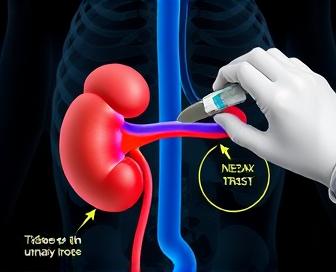
Lasix renograms are typically performed using both the needle and the injector, where a small amount of furosemide (also called Lasix) - an anti-tumor medication commonly used to treat edema and high blood pressure – is injected with 0.5 milligrams of Tc-99m. By highlighting the kidneys' ability to filter and concentrate urine, this combination is advantageous. As the patient is placed on an examination table, a unique camera called 'a gamma camera' takes pictures of how dye moves through their kidneys for several minutes.
Advantages of Lasix Renogram: Non-surgical test specializes in detecting and diagnosing renal problems. Through the provision of detailed information on every kidney's function, doctors can identify potential problems and monitor treatment progress. With a duration of roughly 30-60 minutes, the procedure is relatively quick with few risks and side effects.
Despite its safety features, the Lasix renogram may pose challenges for some individuals. Those who have specific medical conditions, such as severe heart failure (if any), may need to be extra careful before taking this test. You should consult a doctor about any issues before going through with the operation.
Understanding The Lasix Renogram Procedure
Non-invasive nuclear medicine imaging tests, such as the Lasix renogram (diuretic receptacles), are used to detect abnormalities in kidney function. The thorough examination assists doctors in identifying different ailments linked to the kidneys, such as kidney blockage or reflux, and disease development.
Technetium-99m (Tc– 99mm) is a small amount of radioactive material that is injected into the artery in the arm during the procedure. The patient is given Lasix, a diuretic medication that increases urine production through injection. During the filtering and processing of medication by the kidneys, the material is known as Tc-99m, which collects in the tubules near the end of the renal tubular cavity.
After that, a Gamma camera captures several images of the abdomen over an extended period, which helps to track how radioactive material is moving through different organelles such as the kidneys, ureters, and bladder. The images offer a window into the kidneys' blood collection, urine concentration, and response to diuretic stimulation.
During the analysis of test results, special software generates curveS and displays them to show uptake patterns for each kidney in which Tc-99m material is used or excreted. Identifying these curves can aid healthcare providers in considering factors like.
- renal blood flow.
- Kidney function (glomerular filtration rate or GFR)
- Tubular function and concentration ability.
- Uterine permeability.
- Any reluctance, reflux, or other anatomical complications.
In a normal Lasix renogram, the kidneys should gradually increase the uptake of Tc-99m material and excrete it due to diuretic stimulation. Symptoms may include kidney dysfunction, disease progression, or obstruction. They often display patterns or deviations from expected results.
| Normal Lasix Renogram Pattern | A gradual increase in uptake and excretion of Tc-99m material over time, indicating normal kidney function |
|---|---|
| Abnormal Patterns: |
|
By conducting a renogram with the Lasix technique, physicians can examine their patient's kidney anatomy and organ function. The examination of these test outcomes enables physicians to identify various types of renal disease, monitor the development of the disease, and prescribe drugs or therapies that are appropriate for their patients' needs.
What Is A Lasix Renogram?
For example, a lasix (or right) renogram is used to measure kidney function, especially measuring blood flow through the kidneys and also for factors such as glomerular filtration rate (GFR). Through the use of non-invasive techniques, a small amount of specialized dye is introduced into the bloodstream and then transmitted through the kidneys, providing doctors with an opportunity to utilize imaging technology to evaluate their performance.
Technetium-99m, a radioactive substance, is used in conjunction with Lasix (furosemide), also known as flopresemicide. An exception exists, and this combination allows for precise measurement of both renal blood flow (respiratory) and glomerular filtration rate. The kidneys' health is monitored through a series of X-ray scans that take multiple measurements, one after another, using intravenous dye.
Consequently, these images show how quickly the dye is taken up by the kidneys and excreted; they offer valuable information about an individual's health in terms of kidney health and potential risks for disease or damage from conditions such as diabetes or hypertension. To promote the recovery and management of fluid buildup, Lasix is frequently used in acute kidney injury (AKI) diagnosis through a lasix renogram.
Overall, the lasix renogram procedure offers a precise and reliable way for healthcare providers to assess kidney function and make informed decisions about treatment and management plans for patients with kidney-related issues.
How Does It Measure Kidney Function?
During the Lasix renogram, how does renal function track kidney activity with respect to furosemide (the specific drug that controls some of the body's chemicals)? Through the action of this diuretic, doctors can detect if their kidneys are capable of producing enough water and determining whether they can filter waste and concentrate urine.
- During the test, radioactive iodine is injected into the patient's vein in a small amount. The kidneys absorb this substance.
- The intravenous administration of furosemide (Lasix) takes approximately 30 minutes. iodine) the kidneys release and concentrate the absorbed resulting urine.
The radioactive iodine is excreted at different rates by each kidney, and images are captured by a gamma camera placed above the kidneys. These images are analyzed to determine:
- The time required for the kidneys to initiate concentrating urine.
- Their rate of discharge, which includes the excretion of waste products and fluids into the urine.
These findings between kidneys help identify abnormalities or dysfunction. These tests are then expressed in percentage as to the proportion of function exhibited by each kidney compared to its better functioning counterpart, which gives an accurate reading of individual kidney function.
Preparation And Steps Of The Test
A non-invasive Lasix renogram is performed on the kidney to determine the amount of radioisotope that has been taken, as well as the time it takes to wash out and excrete from the body. This test requires a few simple preparations.
- Eating low salt to get the best possible results is recommended for patients who have been undergoing a vitamin C screening for several days before the test.
- Indications of fluid retention: Patients are advised not to drink too much fluid to prevent the radioisotope from being diluted.
- On the day of the test, it is recommended that the patient consume only water and refrain from eating anything beyond midnight until they arrive at the testing facility.
After arriving, patients put on a hospital gown and lay on an x-ray table. The bladder is obstructed during the test by a small catheter that monitors the amount of urine produced.
- A radioisotope (technetium-99m DTPA) through an intravenous line.
- The radioisotope is injected slowly over a few minutes.
- Following the injection, the patient is placed under a gamma camera to record images of their kidneys as the Isotope is taken up and excreted.
| Stage | Description | Duration (minutes) |
|---|---|---|
| Uptake Phase | The radioisotope is absorbed by the kidneys. | 3-5 |
| Washout Phase | The kidneys excrete the radioisotope into urine. | 10-15 |
| Renogram Images | Pictures of kidney function are taken during the washout phase. | Variable (dependent on imaging duration) |
The entire test is 30-40 minutes long. The patient can resume their normal activities right after the procedure, but it is important to drink plenty of fluids as they will help flush out any remaining radioisotopes.
Data Analysis And Interpretation
In this section, we will delve into the process of analyzing and interpreting the data collected during a Lasix renogram procedure. Consequently, it is important to have precise interpretation in order to determine proper kidney function and identify any potential issues that may require further evaluation or treatment.
During the analysis of the renal scan images, special software is utilized to quantify parameters such as time-to-peak (in milliseconds), peak count rate (on an image), and clearance rates. By examining these values, one can gain insight into the kidneys' ability to perform blood filtering and urine concentration as well as excreting waste products.
- The period of time it takes for the radioactive tracer to reach its maximum concentration in the renal parenchyma is referred to as Time-to-peak (TTP). TTP levels that are not normal can be a sign of decreased blood flow or damage to specific parts of the kidney.
- Maximum radiation intensity detected within a given time frame is represented by the peak count rate (PCR). The absence of protein synthesis (PCR) may indicate impairment in kidney function or obstruction in the urinary tract.
- Clearance rates such as effective renal plasma flow (ERPF) are used to determine the ability of the kidneys to remove waste products from the bloodstream and other fluids that pass through them. A decrease in glomerular filtration or nephron damage can be indicative of impaired permeability and flora breakdown (RFP) being present.
A tabular format is commonly used to present the analysis of these parameters for easier comparison and interpretation.
| Parameter | Normal Range | Patient Results |
|---|---|---|
| Time-to-Peak (seconds) | 50-120 | 90 |
| Peak Count Rate (counts/second) | 3000-6000 | 4500 |
| Effective Renal Plasma Flow (mL/min) | 350-550 | 420 |
This is then used to evaluate and interpret the results, depending on individual patient characteristics as well as their medical history and any signs or symptoms of renal dysfunction. For instance, if a patient's time-to-peak is significantly prolonged compared to normal values, it may indicate decreased blood flow to one or both kidneys, potentially due to renal artery stenosis or occlusion.
Importance Of Accurate Kidney Function Assessment
The removal of toxins, fluids, and waste from the body is one of the many important functions of the kidneys that are essential for maintaining good health. Consequently, the accurate evaluation of kidney function is essential for diagnosing and treating various kidney-related diseases. Through this examination, medical professionals can identify the earliest signs of kidney damage or dysfunction, which may result in chronic diseases if left untreated.
Accurate testing of kidney function enables doctors to monitor changes over time, evaluate the effectiveness of treatment plans, and modify medication as necessary to prevent potential complications. To illustrate, it aids in determining the appropriate amount of dialysis for individuals with terminal renal disease or prescribes the most effective medication regimen for those with chronic kidney disease (CKD). In addition, the accuracy of assessment enables healthcare professionals to identify individuals at high risk for developing CKD, allowing for early intervention to slow down the disease.
The outcome can be poor due to inaccurate or incomplete assessments, which may lead to misdiagnosis, inadequate treatment, and poor health outcomes. If kidney function is not accurately assessed, there is a risk of inadequate fluid removal during dialysis, which can lead to life-threatening complications like fluid oversufficiency. Conversely, an overestimation could result in unnecessary interventions or medication modifications that may worsen the condition.
The assessment of kidney function is not solely for patient care. This information is used to inform public health policies and research efforts aimed at preventing CKD, developing better treatment options, and improving the quality of life for individuals with kidney disease. By educating patients about the importance of regularly testing their kidneys, healthcare providers can help patients become more proactive in taking steps to maintain their renal health.
The accurate determination of kidney function is crucial for providing personalized treatment, preventing complications, and contributing to our understanding of chronic kidney diseases. Therefore, it is still a fundamental element of the practice of nephrology and public health initiatives that aim to enhance patient outcomes and quality of life.
We recommend you read it
Many people use Lasix as a diuretic. Its side effects are similar to those of other medications.
- Study the impact of Lasix on lymphedema.
- Discover the indications of acute kidney injury (AKI) linked to Lasix use by examining the symptoms.
- Discover information on the process of changing Lasix dosages from IV to oral by mouth for optimal results.