


Nuclear Medicine Renal Scan with Furosemide (Lasix)
Search:
ADS:

Nuclear Medicine Renal Scan with Furosemide (Lasix)
To evaluate the health of the kidneys and urinary tract, a nuclear medicine renal scan is conducted without intrusion. a small amount of radioactive substance which is then fed into the body and tracked through by.
In this procedure, furosemide (Lasix) may be given to increase urine production and improve image clarity. Medics can identify various health issues, including kidney damage caused by an ailment, reflux, or other diseases or injuries, through the use of this test.
Acknowledging the process and preparing beforehand can alleviate anxiety and ensure success. We would like to go over the basics of nuclear medicine renal scans with Lasix, including what is involved, necessary preparations, and how these are likely to impact outcomes.
By filtering waste products, regulating electrolytes, and producing hormones that regulate red blood cells and maintain healthy blood pressure, kidneys are essential for maintaining good health. The health of patients can be significantly affected by any impairments to their kidney function.
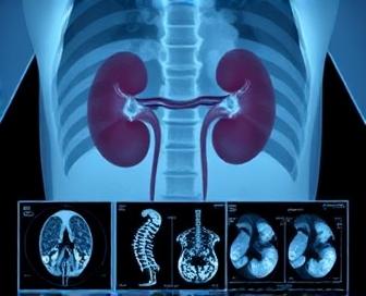
The utilization of nuclear medicine renal scans is beneficial in identifying and tracking kidney diseases. Detailed images of kidney blood flow and urine buildup are provided by this test to doctors, which helps them identify potential issues and develop appropriate treatment plans.
Nm Renal Scan With Lasix Explained
The kidneys' structure, function, and surrounding structures are scrutinized through a diagnostic test known as recurrent renal scans. This tool aids doctors in identifying kidney-related issues such as stones, tumors (endoscopals), infections, or blockages. To visualize the kidneys, a gamma camera produces images of the renal scan using Nm (nuclear medicine), which uses small amounts of radioactive materials, typically technetium-99m, that are injected into the arteries.
A diuretic drug called Lasix (furosemide) is used as a test, which can enhance urine production. By using Lasix, an Nm renal scan can reveal more accurate and detailed information about the functioning of the kidneys than an average Nmm renal scanner.
This test may demand that patients:
| Fluid restriction | Limit fluid intake to a minimum, usually 8 hours before the test. |
| Medication adjustments | If taking diuretics or other medications that can interfere with the test's results, they may need to be adjusted or temporarily stopped under medical guidance. |
| Fasting | In most cases, patients are asked not to eat for at least 4 hours before the scan and avoid consuming any fluids except water within a few hours prior to the test. |
In the procedure, an IV line is inserted into another vein (usually inside our arm). During the short interval (usually around 30 seconds), the radioactive material and Lasix are injected through this line. The administration of medication can result in a short-lived stinging sensation or heightened body temperature during the injection.
Several minutes after the injection, a Gamma camera takes pictures of the kidneys. The pictures will exhibit the kidneys' efficiency, encompassing blood circulation, urine production amounting to 90% of normal fluid volume, and any potential obstructions or damage within the structures of the organ.
After the assessment, individuals can promptly resume their usual eating and engaging in activities. The results from an Nm renal scan with Lasix typically take a few hours to be analyzed by a radiologist who will review the images and provide a detailed report outlining any abnormalities found in the kidneys' functioning or structure.
Understanding the Procedure
The use of Lasix in a renal scan is primarily for diagnosing kidney function, with sensitivity to potential abnormalities like scar tissue, tumors, or blockages being taken into account. It's performed after an injection of a small amount of radioactive material called technetium 99m and the diuretic Lasix. During the procedure, doctors are given crucial information to diagnose and treat different types of kidney disease.
Exercise is necessary: Those who are scheduled to take this examination usually require around 4-6 hours of fasting and abstain from caffeine or fluids for 2-3 hours before the test. It is possible that they will be instructed to avoid wearing items made of metal that could potentially harm scan images.
It takes approximately half an hour for scanning to happen, during which the patient is placed on an x-ray table and has one of its gamma cameras capture images of their kidneys as they function. The injected Lasix helps increase urine production, allowing doctors to assess how well the kidneys filter waste from the bloodstream.
| Step | Description |
|---|---|
| 1 | Injection of technetium 99m and Lasix |
| 2 | Patient lies on x-ray table for gamma camera imaging |
| 3 | (5-15 minutes)Lasix increases urine production for assessment |
| 4 | Gamma camera captures images of kidneys functioning |
| 5 | (20-40 minutes)Additional imaging may be taken to gather more information |
| 6 | Results are analyzed by a radiologist and communicated to the ordering physician |
Doctors can gain valuable insights into kidney function, blood circulation, and structure from the scan results. They can also identify areas of reduced blood flow or impaired filtration capacity, which may help diagnose conditions such as chronic kidney disease in those with active bladder infections, acute kidney injury that causes obstructions in the urinary system, and tuberculosis (throat spasms during the delivery of intravenous fluids through incomplete drainage systems) to specific sites of increased perfusion.
Preparing for the Test
The preparation for a nuclear medicine renal scan with Lasix demands significant effort. The examination necessitates the consumption of a radioactive contrast agent, Lasix (furosemide), and supplementary amounts of an insignificant amount of radioactivity.
- You should arrive early at the nuclear medicine department on the day of the test, as directed by your doctor, usually an hour before the scheduled start time. This affords ample time for preparation and registration.
- You will be instructed to change into a hospital gown or comfortable clothing without metal fasteners or zippers, which may interfere with the imaging process.
- To administer the radioactive contrast agent and material, a nurse or technician will insert an IV line into your arm (or your hand) to inject it.
Your doctor may recommend particular dietary guidelines for individuals who want a renal scan using Lasix in nuclear medicine. All-in-all, you should do:.
- The use of foods that contain high potassium should not be done for at least 24 hours before the test, as it could affect how your body reacts to Lasix.
- Avoid taking any potassium-containing drugs for a period of 2 days, as long as it is within reach before the test. Your doctor will give you specific instructions on what medication to take during this time.
Pregnancy and breastfeeding require you to discuss your situation with a healthcare professional before having the test. These cases may require the implementation of specific measures to decrease radiation exposure and guarantee a safe checkup for both mother, child, and caregiver.
The Role of Lasix in the Scan
Lasix is a vital component of the Nm Renal Scan, serving as an essential treatment. The function of this is not just to improve the quality of the scan; it greatly enhances the diagnostic procedure.
- By acting as a diuretic, Lasix triggers the kidneys to produce more urine, and it temporarily increases blood flow through the renal vessels.
- This boost in blood flow also improves the viewing of these organs' function during a scan, meaning clinicians can better assess perfusion as well as those requiring filtering or excretion.
- By raising the output of urine, contrast agents utilized during a scan can be eliminated from the bloodstream, which lowers the likelihood of nephrotoxicity and other complications.
- The diuretic properties of Lasix enable the kidneys to eliminate waste products and fluids, which can be a risk factor for many medical conditions or medications. This clearance is essential for maintaining normal kidney function during the scan.
Initially intended to enhance contrast, Lasix has been extensively utilized in the Nm Renal Scan. This is important because of its diuretic effects, which help to optimize the scan quality, ensure patient safety, and provide diagnostic information about renal perfusion and function with high accuracy.
Interpreting the Results
The use of Lasix in an Nm renal scan can reveal important details about kidney structure, function, and blood circulation. These images are subject to careful scrutiny by a nuclear medicine specialist or radiologist who examines them closely to determine whether kidneys are in proper working order and identify any abnormalities.
- Normal Kidney Function: In a normal examination, the results show both kidneys functioning at expected levels, with symmetric uptake of radionuclides and rapid clearance after Lasix administration. Healthy kidneys are responsible for enabling efficient excretion and waste elimination.
- Kidney Scarring or Fibrosis: Chronic damage to the kidneys can occur due to various reasons, including scarring of one or both kidney(s) - for example, by hypertension, diabetes (one blood cell per capsule), and glomerulonephritis (thrombosoa). The presence and extent of scarring help assess the stage of kidney disease.
- Renal artery stenosis is caused by the narrowing of the renal arteries, which can hinder the flow of blood to the kidneys and potentially impair their function. In the given scenario, the scan may indicate either asymmetrical uptake or delayed clearance due to decreased perfusion in the affected kidney.
- Kidney cysts or tumors: The presence of benign or malignant kidney stones, tumors, and other lesions can lead to increased radionuclide uptake in focal areas. The diagnosis and treatment plan often require follow-up scans and biopsies.
- The dilatation of the renal pelvis and calyces occurs when urine is returned to the collecting system due to obstruction or reflux, which is a sign of hydronephrosis. Retention of radionuclides and delayed drainage can result in characteristic imaging patterns.
Comprehending the results of Nm renal scans with Lasix requires knowledge of normal kidney physiology and anatomy. Diagnosis of different types of kidney disease is often carried out by physicians who identify anomalies in the expected results, as well as assess the severity of the disease and develop appropriate treatment plans to improve patient outcomes.
We recommend you read it
Our page Does Lasix Make You Pee? provides in-depth information on the impact of Lasix on urination. You can find detailed information on the impact of Lasix on your body in this article.
For those who are concerned about possible side effects, read our page: "Is it true that Lasix raises the levels of b vitamins and creatinine?" We explain the impact of Lasix on kidney function and how it impacts your health.
Additionally, Lasix's effect on lymphedema is noteworthy. Learn more by reading our article Does Lasix Help Lymphedema? By analyzing the possible uses and drawbacks of Lasix for this illness, you can trust our experts to provide an accurate explanation.