


Nuclear Medicine Renal Scan with Lasix Interpretation for Efficient Kidney Function Analysis
Search:
ADS:

Nuclear Medicine Renal Scan with Lasix Interpretation for Efficient Kidney Function Analysis
To accurately evaluate kidney function and detect any potential issues, a renal scan is frequently used in nuclear medicine. One common method involves administering the diuretic lasix to enhance image quality and provide additional insights into kidney performance. By utilizing nuclear medicine and lasix, this article provides an overview of the techniques and interpretations used in a renal scan with laspicion, which aid healthcare professionals in optimizing their analysis of kidney function and making informed treatment decisions.
The kidneys selectively absorb small amounts of radioactive material, known as radiotracers, during a renal scan. Spectacular cameras can detect the emission of GammA-rays by the patient's body during the processing of these tracers. This enables the production of comprehensive visuals of the kidney's structure and function.
This procedure may involve the use of lasix (furosemide), which is another option. Initially, it boosts urine production, which enhances the detection of renal parenchyma and cortical structures on the scan. Lastly, Lasix assists in distinguishing between normal and abnormal kidney functions by emphasizing functional differences. Finally, the diuretic impact enables more precise evaluations of kidney size, shape, and perfusion.
Through the use of expert interpretation, healthcare professionals can obtain a comprehensive understanding and early detection of potential issues, while also monitoring treatment responses. This paper will discuss the various methods utilized in nuclear medicine, including complex processes and tips for optimal scan analysis when performing renal scans with lasix.
The initial picture depicts a Nuclear Medicine Renal Scan with Lasix.
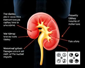
Enhanced view: This image shows the improved view of kidney structures after use with lasix in a renal scan taken during nuclear medicine.
Nuclear Medicine Renal Scan
The use of radioactive tracers through a nuclear medicine renal scan is aimed at testing kidney function, making the procedure non-invasive. By absorption by the kidneys, specialized cameras can detect the tracers and provide detailed visual assessments of various organs and functions. Diagnosis of various renal disorders can be achieved through this thorough examination, encompassing acute kidney damage and chronic conditions such as kidney disease, hydronephrosia, or vascular impairment.
To get better images, patients may be given diuretic medications like Lasix by mouth to prepare for the test, and they usually go without food for 4-6 hours before testing. A gamma camera captures images of their kidneys while they are placed under them during the procedure, where tracer beams are built up and excreted.
Analyzing the scan results using parameters such as renal uptake, peak activity rate, time-to-peak times (such as bedside fever), and washout rates is carried out by radiologists. These are useful measures for monitoring renal activity; the ability to detect defects in peripheral blood flow or filtration; and the detection of potential obstructions such as kidney stones, tumors, or other pathogens. With the Lasix diuretic, images can be visualized more clearly by promoting the rapid removal of radioactive tracer channels.
Nuclear medicine renal scan has several advantages over other imaging methods. The early detection of early signs of kidney disease and measurement enable it to be used for the diagnosis and management of patients with chronic kidney failure or those at high risk for renal impairment caused by obstructive uropathy.
Understanding the Procedure and Imaging Techniques
Nuclear medicine renal scans with Lasix administration are a diagnostic tool used to assess kidney function, particularly in the context of hypertension. Injecting technetium-99m (Tc–98M), a small amount of radioactive substances into. Once absorbed by the kidneys, it is concentrated in this substance within its own structure in the renal parenchyma and then released into the urine.
After receiving Tc-99m, furosemide (also known as Lasix) is given intravenously to stimulate urinary flow and cause concentration shifts in the kidney. It also allows radiologists to better visualize the structural features of a person's kidney and helps assess function, especially when there is potential renal (potential) disease or damage.
- The typical duration of the procedure is roughly one hour, with additional time allocated for patient preparation.
- The kidneys are photographed by a gamma camera at all stages: before, during, and after the procedure of Lasix, as well as during and following the diuretic effect.
- The radiologist's interpretation of these images involves taking into account factors like renal uptake and washout rates, cortical thickness in the eyes, and the relative function between the two kidneys.
- The use of computer analysis software can enhance image quality and facilitate quantitative measurements of kidney function.
Nuclear medicine renal scans with Lasix are used to interpret them, and they often focus on abnormalities in the structure or function of a kidney that could be responsible for hypertension. Commonly discovered indications comprise:
- In areas of kidney damage, including scarring caused by diabetes or high blood pressure, there is a decrease in uptake or delayed washout.
- Distinctive functioning of the two kidneys, indicating potential obstruction or disease in one.
- Larger-than-normal kidneys due to chronic hypertension, indicating long-term damage and scarring within the organ.
- Enlargement or ectopic kidneys caused by congenital abnormalities or trauma.
Radiologists and clinicians must possess a thorough understanding of the nuclear medicine renal scan with Lasix interpretation techniques to diagnose kidney-related disorders with precision and develop treatment plans.
Distinguishing Normal from Abnormal Findings
Using Lasix diuresis to differentiate between normal and abnormal renal scan results, we will now discuss how this can be identified and interpreted in this section. A Lasix stress test that closely reflects the expected changes in renal function and perfusion is essential for accurate diagnosis.
A healthy kidney may experience rapid diuresis and rapid washout due to the use of furosemide (Lasix) in both lungs. The activity levels rise to peak early but then decrease to normal within 10-15 minutes of injection. The DRF should be symmetrical, with no discernible differences between the two sides.
Abnormal Findings: Conversely, abnormal scans may reveal delayed or absent diuresis, indicating impaired kidney function. This can manifest as prolonged activity retention in one or both kidneys, often accompanied by asymmetry of DRF. Other potential indicators of pathology include: 1) persistent activity accumulation on the renal pelvis or calyces (> 30 minutes post-injection), suggestive of obstruction or stenosis; 2) focal defects in perfusion or function, potentially representing areas of scar tissue, infarction, or inflammatory lesions.
To accurately interpret these findings and differentiate between benign conditions like hypertrophy or ectasia, which can mimic abnormal results, and true pathology, it is important for the radiologist to be well-versed in this area. By utilizing the findings from the kidney scan and providing clinical information, such as medical history or symptoms, along with laboratory tests to guide management, patients can receive a comprehensive diagnosis that can be followed in the future.
Interpreting Lasix Stimulated Images Accurately
To accurately interpret the images produced by Lasix-stimulated imaging in nuclear medicine, one must have a good understanding of the physiology involved. By causing diuresis, Lasix (furosemide) stimulates urine production and affects the kidney's function, leading to the uptake and excretion of radiotracers. This leads to the development of useful data on regional blood flow and filtration capacity in every kidney through the resulting images, as well as tubular function.
Renal function must be evaluated when interpreting these stimulated images. This is because the scan can only detect defects or abnormalities in renal function, so how sensitive is it to the degree of diuresis produced by Lasix (predominantly from edema)? When the kidney is functioning properly, it will concentrate more of the radiotracer and show increased uptake; when it is not, the uptake may be delayed or reduced, possibly due to reduced perfusion (leading to tubular dysfunction) and/or tubal edema.
The images captured during Lasix stimulation reveal that Uptake Defects can occur in areas with low radiotracer concentration, Persistent Trapping can result in delayed clearance or retention of the radio tracer in a specific area, and Heterogeneity can cause uneven distribution of activity within kidneys. The manifestations can be a sign of various diseases, such as renovascular disease, glomerular disease (in the absence of blood), tubulointerstitial disease, or obstructionive nephropathy.
It is important to take into account the patient's medical history and clinical presentation when interpreting images with Lasix-stimulated images, in addition to the distribution patterns seen through radiotracers. By analyzing the visual cues of their individual structures and movements, each kidney is evaluated for its overall function as well as cortical and medullary uptake, including any abnormalities or asymmetries. Nuclear medicine specialists can use this visual assessment in conjunction with knowledge of the effects of Lasix-induced diuresis on renal physiology, which they believe is useful for clinicians to understand the causes of renal dysfunction.
Case Studies for Better Understanding
The use of Lasix during a nuclear medicine renal scan is demonstrated through specific examples in this section, providing an overview of the application of different interpretation techniques. Through the use of real-life scenarios, readers can gain a better understanding of how imaging findings are utilized in diagnosing and managing underlying renal disorders.
Case Study 1 involved a 65-year-old woman who had pain and hematiolysis on her flanks. She had a nuclear medicine renal scan, which revealed she was suffering from acute (potential) formative pyelonephritis and had reduced function in her right kidney. The patient was given antibiotics and made a full recovery within two weeks. During the case, the scan results accurately pinpointed where and how much of the infection was present at that time, making it easier to treat with specific measures.
On the other hand, Case Study 2 centered on a 40-year-old man who had been diagnosed with renal cell carcinoma and had undergone invasive surgery such as partial nephrectomy several years earlier. Following the surgical removal of a small mass from his left kidney, he underwent another nuclear medicine renal scan to evaluate the function of the remaining kidney. No significant impairment was detected in the scan, indicating that the patient's remaining kidney could function normally and meet all its physiological demands.
In Case Study 3, extrarenal factors are considered when interpreting nuclear medicine renal scans. The 55-year-old diabetic patient experienced deteriorating peripheral edema and hypertension. While her scan revealed only a very mild case of bilateral hydronephrosis, the more serious later diagnosis was that she had developed severe peripheral artery disease.
These case studies demonstrate that nuclear medicine renal scans with Lasix can provide useful diagnostic information for a variety of renal disorders and conditions that may affect kidney function. These real-world applications enable clinicians to interpret scan results and make informed treatment decisions for their patients.
We recommend you read it
Our informative materials on Lasix's effects are outlined below:
- Does Lasix Increase BUN and creatinine? - Get answers on how this diuretic may impact your kidney function.
- Can Lasix alleviate lymphedema? Gain insight into the potential benefits of using this antidote.
- Does Lasix contain sulfa? (Read more about) What is the toxicity of SFAC in this medicine and should necessitate SLS for someone with SSFE?