


Renal Lasix Scan in Radiology: An Overview of Diagnostic Benefits and Technique
Search:
ADS:

Renal Lasix Scan in Radiology: An Overview of Diagnostic Benefits and Technique
The renal Lasix scan is a groundbreaking non-invasive diagnostic imaging method in radiology. Doctors can diagnose various kidney and urinary tract disorders by utilizing this innovative technique. With the aid of high-powered imaging technologies and high contrast agents, the renal Lasix scan provides unparalleled knowledge of kidney physiology and pathophysiological processes.
A renal Lasix scan is primarily used to evaluate kidney function, with the test being performed to determine how quickly the kidneys filter waste products from the blood. This involves injecting a small amount of furosemide, which is also called Lasix, containing the active ingredient that diurestics work. With its effect, the drug triggers the kidneys to excrete more urine; radiologists can observe the flow of contrast material through the renal parenchyma and urinary collecting system.
These detailed pictures offer doctors a comprehensive understanding of the structure and function of your kidneys, helping to identify what'd happen in your body abnormally (child urine), cysts or blockages, etc. The method is particularly advantageous for identifying illnesses like chronic kidney disease, nephrotic syndrome, and hydronophrosis. Moreover, the kidney Lasix scan provides an opportunity to assess whether treatment strategies aimed at improving renal function are effective.
An examination table is typically used to hold patients while they are positioned for the procedure, which involves the placement of an intravenous catheter in a vein. The patient is subjected to a Lasix injection before being placed under the scanner, and images are processed using computeducytomography (CT) or magnetic resonance imaging (MRI) techniques. The whole thing typically takes 30 to 60 minutes in duration.
Several advantages of the renal Lasix scan compare to conventional diagnostic procedures. This may aid in distinguishing between chronic kidney disease and acute kidney injury, which are often unidentifiable based on clinical signs. In addition, the test is non-invasive -- meaning it does not pose any risks as would invasive tests such as a kidney biopsy.
While a renal Lasix scan is now commonly used for radiological diagnosis, it should be noted that the test should only be performed under the supervision of specialized medical professionals. Special care may be required for pregnant women and certain medical conditions, or alternative imaging modalities may also be available.
A groundbreaking new approach to imaging kidney function is the Lasix scan. Through this groundbreaking innovation, radiologists can now diagnose and treat complex kidney conditions with greater precision, thanks to its unparalleled understanding of their function.
Renal Lasix Scan Radiology Explained
To assess kidney function, abnormalities, and medication response, a non-invasive imaging test called LASix is performed. To enhance the visualization of the kidneys on images, radiography (x-ray) is combined with an intravenous injection of furosemide (LaSix) in this diagnostic procedure. The assessment enables clinicians to determine whether individuals with chronic kidney disease, hydronephrosis, or obstructive renalpathies are affected by the test.
Evaluating preparation: It is customary for patients to consume a significant amount of water before the assessment and abstain from consuming caffeine or diuretics in the hours before their evaluation. Then, a vein is used to administer furosemide after an intravenous opening. An x-ray table with the patient's legs elevated is typically used for imaging purposes.
The protocol entails taking X-rays right after injecting Lasix and at regular intervals for 30 minutes to 1 hour. These pictures show the kidneys in varying stages of operation; radiologists can determine their appearance, size, shape, and location. The acquisition of images can be achieved using either DR or CR technology.
An expert in radiology analyzes the images obtained by a trained technician to detect any deviations, such as dilatations of their collection's structure, areas of reduced function, or structural changes. The kidney's performance is monitored over time using test results, as well as previous studies and normal references (if available).
The kidney's function and structure can be accurately assessed through the safe and effective use of a renal Lasix scan, which clinicians are trained in using to make informed decisions about patient care. Patients can learn about the principles of this examination by following it, which will assist them in preparing and anticipating their test.
Understanding the Purpose of a Renal Lasix Scan
Diagnosis using a renal Lasix scan, also called 'diuretic radionuclide renography,' is an imaging test used to provide valuable information about kidney function and structure. To determine the effectiveness of each kidney, a non-invasive procedure is used that uses diluted radioactive material and furosemide (Lasix) to measure renal function.
The patient drinks a solution containing 10% technetium-99m, preferably sourced from. Through the absorption of this into the kidneys, they can be captured on camera by a dedicated technician. Lasix medication temporarily boosts urine production, allowing for better monitoring of kidney function.
To identify urinary tract abnormalities or obstructions, a renal Lasix scan is typically performed.
| Abnormality | Description |
|---|---|
| Kidney obstruction | A blockage that prevents urine from flowing properly through the kidney. |
| Nephropathy | Damage to the kidneys due to various causes, such as diabetes or high blood pressure. |
| Urinary tract reflux | A condition where urine flows back into the ureters from the bladder. |
| Vesicoureteral reflux | A specific type of urinary tract reflux affecting the kidneys and urinary tract. |
| Pyelonephritis | An infection that occurs in the kidneys, often caused by bacteria entering through the ureters. |
By utilizing the scan, healthcare providers can:
- Assess the condition of a kidney;
- Identify the UTI;
- Check for any breakthroughs in the treatment of different types of kidney diseases.
By using a renal Lasix scan, it is possible to accurately identify and manage various kidney disorders that are not commonly understood without the use of other methods. By doing this, healthcare providers can detect potential issues at a glance, enabling them to effectively prevent any complications from occurring later.
Preparation for Your Renal Lasix Scan Appointment
A few things can be done beforehand to ensure your renal Lasix scan appointment goes smoothly. You will experience less stress and anxiety during the exam if you are adequately prepared. This section outlines the key preparation steps you should follow prior to your renal Lasix scan.
To start with, ensure that you have a complete record of the date, time, location, and any specific instructions from your radiology department or imaging center where your exam will be held. Don't forget to ask for information on parking, accessibility for those with mobility impairments, and what to expect when you arrive. Centers may offer a detailed guide, so it's possible to ask for one.
Subsequently, recollect any specific preparation guidelines provided by the radiology department or your physician. These might include:
- hours without food and fluids before the exam.
- Relaxed, light clothing that allows for easy movement.
- Removed jewelry, glasses, or metal objects.
- Arriving 15-30 minutes before your scheduled appointment time.
- Provide medical records, insurance cards, and identification.
If you have a pacemaker, implantable cardioverter-defibrillator (ICD), or other implanted device, please let the radiology department know in advance of your examination so they can take appropriate steps during the exam.
Ultimately, get ready mentally for the operation. Remember that renal Lasix scans are painless and non-invasive. Maintain a calm and peaceful approach while conversing with your physician.
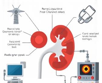
Radiology Techniques Used in a Renal Lasix Scan
The kidneys are imaged in great detail during a Lasix scan, which is obtained through the use of various radiology techniques. With the aid of these techniques, physicians can examine a kidney's condition accurately, identify any abnormalities, and monitor treatment progress. Among the most important methods used are intravenous pyelography (IVP), computed tomography (CT), magnetic resonance imaging (MRI), and single photon emission computed tomography (SPECT). Each approach provides distinct advantages in assessing renal structures and functions.
The IVP procedure involves injecting a contrast agent into the vended arms. The X-rays are used to visualize the urinary tract as it moves through the kidneys, which can help identify any obstructions or abnormalities. Using low-radiation X-rays and computer algorithms, CT scans provide high-resolution cross-sectional images of the kidneys, which can be used to gain insight into their shape, size, and blood circulation.
The use of high-resolution images of soft tissues like kidneys in MRI is possible due to the absence of radiation exposure thanks to strong magnetic fields and radiofrequency pulses. Diagnosing conditions affecting kidney parenchyma or surrounding structures is made easier with this technique. Through the use of SPECT scans, nuclear medicine and CT imaging can be combined to reveal the distribution of radiotracers throughout the body, allowing doctors to assess kidney function and identify areas where oxygen consumption is reduced.
The choice of radiology technique used for a renal Lasix scan is influenced by the patient's medical history and individual needs. A thorough examination may involve combining various methods to achieve a more precise diagnosis. With the help of these newer imaging techniques, healthcare providers can more precisely assess and manage kidney-related illnesses, resulting in better treatment outcomes and care that patients expect from their physicians.
Interpreting Renal Lasix Scan Results
This section will provide an overview of the renal Lasix scan process. Healthcare professionals must be informed about these findings to make appropriate decisions regarding patient care. An understanding scan can help identify potential kidney disease and provide treatment options.
Assessing Kidney Function
- This parameter is used to measure the rate at which blood flows into the kidneys, known as renal blood flow (RBF). The mean RBF is generally around 550-1100 ml/min. Abnormal findings may indicate kidney damage or disease.
- Effective renal plasma flow (ERPF) or ERPF is the measure of the quantity of plasma that passes through the glomeruli at a given time. Normal levels of ERPF are 115–140 mL/min. Severe hypertension, renovascular disease, or chronic kidney disease are frequently linked to decreased values.
- GFR (Glomerular Filtration Rate): GFR is a key indicator of overall renal function and represents the rate at which the kidneys filter waste products from blood into urine. Normal adult GFR ranges are around 90-120 mL/min/1.73m2 body surface area.
Lasix (Furosemide) Response
This is measured by the Lasix response, which measures how well the kidneys work to concentrate urine when subjected to diuretic stimulation. If the patient's response is normal, it may be due to nephrotoxicity or chronic kidney disease; if it is suboptimal, its response may not necessarily reflect intact tubular function and a reserve of specialized ocellions known as refractorius.
Pattern Recognition
Interpreting renal Lasix scan results requires pattern recognition across multiple parameters. For example,
- The presence of low RBF and ERPF, in combination with a normal GFR, may indicate severe hypertension or serious renovascular disease.
- Symmetrically reduced renal function in both kidneys may be a feature of chronic liver disease or other conditions, while contrasting findings might be present in those with either type of polycystic kidney disease (RnCd) or renovascular disease.
Limitations and Future Directions
The kidney function and structure can be understood through the use of a renal Lasix scan, but there are limitations to this method. For example,
- This test may be unsafe in individuals with high levels of severe azotemia (higher blood urea nitrogen levels) or acute kidney injury, as it could potentially cause harm from contrast agents.
- The diagnosis may be invalid if there is a falsified positive result in cases of renovascular disease or chronic kidney disease, necessitating additional diagnostic testing with other methods like renal Doppler ultrasound or CT angiography.
Research is being conducted to address these limitations and improve the accuracy of interpretation, with a particular emphasis on refining imaging protocols, creating new contrast agents, and exploring machine learning for improved pattern recognition capabilities in renal Lasix scans.
Post-Scan Care and Recovery Tips
Following a successful renal Lasix scan, it is important to carefully follow post-scan care instructions to ensure proper recovery and minimize the risk of complications. Depending on your individual circumstances, your radiologists or physician will provide you with tailored advice, but there are some general tips to keep in mind.
- Relieve your body: Don't engage in vigorous activity for more than a day after the scan, as excessive physical activity can intensify any discomfort or negative side effects. Give yourself enough rest and do gentle stretching exercises instead.
- Drinking a substantial amount of water throughout the day can help flush away contrast agents from the blood and other fluids used during the scan. Increased urine output is a common result, but it can still occur.
- Pain management: If you experience pain or discomfort, over-the-counter pain relievers like acetaminophen (Tylenol) or ibuprofen (Advil, Motrin) can help alleviate symptoms. Follow the recommended dosage instructions and seek medical advice from your doctor.
- Monitoring for side effects: Keep an eye out for signs of allergic reactions, such as hives, itching, swelling, or difficulty breathing. Immediately seek medical attention if you exhibit any unsettling symptoms.
- Make arrangements for follow-up appointments: Your doctor may arrange follow-up assessments to evaluate the efficacy of treatment and keep track of your progress. Take care of these appointments with great care.
By following these post-scan care tips and adhering to your individualized instructions, you can promote a smooth recovery and achieve the best possible outcomes from your renal Lasix scan.
We recommend you read it
It is important to be aware of the possible side effects of Lasix if you are considering using it for your health condition. Hyperkalemia and hypokalemia are two potential negative consequences of consuming Lasix.
- Read more: Can Lasix cause hypokalemia or hyperkalemia?
- Hypernatremia is a potential consequence of taking Lasix.
- Discover if Lasix triggers hypernatremia.
- Know the correct dilution procedure for administering Lasix via IV push.
- Get informed about should you dilute Lasix iv push.