


Mag3 Renal Scan with Lasix Explained
Search:
ADS:

MAG3 Renal Scan with Lasix Explained
To identify kidney abnormalities or urinary tract abnormalities, a physician may conduct MAG3 renal scans to assess the body's overall health. By injecting a small amount of radioactive substance, MAG3, into safty or an ineluctable stream of blood into the patient'S bloodstream, it is possible for healthcare providers to examine their structure and function using special cameras.
When used with furosemide, Lasix improves accuracy by allowing the kidneys to eliminate excess fluid more quickly during testing. Identifying different types of kidney dysfunction can aid in making informed treatment decisions. MAG3 renal scans with Lasix are described, and the patient's experience is clear.
Reasons for Concerning Kidney Function.
In addition to their important role in maintaining good health, the kidneys also act as a waste product filter and electrolyte iron supplier (which helps control body weight) as well as an anti-platelet microcephaly. Acute kidney failure can result in various complications, such as chronic kidney disease (CKD), acute kidney injury (AKI), and end-stage renal disease necessitating dialysis or transplantation.
The prevention and early treatment of kidney issues can lead to a decrease in long-term side effects. A MAG3 renal scan with Lasix is an essential tool in diagnosing and managing various kidney disorders, providing valuable insights into kidney structure and function that guide therapeutic decisions and improve patient outcomes.
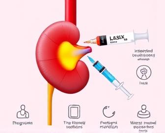
Patients undergo a MAG3 renal scan procedure that begins with drinking small amounts of water to fill their bladder, which is then emptied before the test. MAG3 solution is administered through the use of a catheter into the artery in the arm over 3-5 minutes at varying speeds.
Upon receiving the injection, patients will sit on an examination table while being photographed using a visible light spectrum (gamma camera) to detect and interpret radiation from MAG3 as it builds up in the kidneys. During the scan or shortly before it, Lasix is given to enable rapid urination without any extra fluid accumulation.
The images analyzed reveal precise information about the kidney's structure, function, and blood flow patterns, making it possible for doctors to identify abnormalities like scars from previous infections, blockages, or tumors. Rather than simply examining these images, clinicians can use other patient data to determine the cause of kidney dysfunction and develop an appropriate treatment plan.
MAG3 Renal Scan with Lasix Explained
During a nuclear medicine test, MAG3 renal scan with Lasix is performed to identify kidney function problems like obstruction or scarring. This non-invasive exam can give doctors valuable information about the kidneys' working condition and help diagnose potential problems.
During a short-term outpatient procedure, MAG3 (magnetium-99m) is introduced into the patient's vein as if passing through ice and then injected back into the bloodstream for re-encephalography or to give them symptom relief. The kidneys eliminate the substance promptly, facilitating precise monitoring of blood circulation and function in both kidneys.
The use of furosemide, also known as Lasix, during the scan is to stimulate urine production and improve imaging accuracy by administering it intravenously. By using a combination of the MAG3 Renogram and Lasix injection, doctors can evaluate kidney function in different situations, such as those where blood flow is considered normal or when obstruction occurs to perfusion.
Images taken at different stages following the injection are used to present the test results, which indicate the condition of each kidney and any potential blockages or scars. Through the examination of these images, doctors can identify potential conditions such as pyelonepine disease (which is caused by inflammation), kidney stones, tumors, vascular disease, or congenital anomalies and determine the appropriate treatment for them.
Alternative therapies for managing MAG3 scan abnormalities include medications to manage symptoms, lifestyle changes such as cutting back smoking and hypertension; surgical treatments; or more conventional interventional procedures like nephrostomy tubes. Occasionally, additional diagnostic procedures like CT scans, ultrasound treatments, or biopsies are suggested to obtain more information before making a final decision.
Individuals are advised to adhere to any specific guidance provided by their physician before and after the examination, such as any medication restrictions or changes in fluid quantity. The role of Lasix in the assessment of kidney function and its use can be aided by understanding what a MAG3 renal scan is and how it can assist with patient preparation for this diagnostic tool, as well as working with their physician or other healthcare providers to identify potential problems.
Diagnostic Purpose and Process
To assess kidney function, structure, and blood flow, Lasix performs a type of MAG3 renal scan. This procedure is used as a diagnostic step to determine whether the kidneys are capable of centring urine and excreting waste products. Diagnosis is also aided by it, which covers conditions such as chronic kidney disease (CKD), hydronephrosia, renovascular hypertension, and some forms of glomerulonephritis.
A radioactive substance, technetium-99m MAG3, is usually given intravenously as an early indication for the initiation of the renal scan process. Absorption of this agent into the kidneys enables imaging devices to visualize them. Following the initial injection, patients consume Lasix (furosemide), a diuretic that stimulates urine production.
The kidneys react to the diuretic and process the injected agent, which is then captured in images using single photon emission computed tomography (SPECT) or planar imaging techniques. The kidney's function can be accurately monitored through these images, which exhibit details about blood flow, radiotracer uptake, and excretion patterns. The entire procedure usually takes 30-60 minutes.
A healthcare provider interprets the scan results to identify any abnormalities in kidney structure or function. In the case of a MAG3 renal scan, it is possible to detect delayed tracer uptake or retention, which could indicate impaired kidney function due to conditions like chronic kidney disease (CKD) or renovascular disease. Comparisons with previous scans are sometimes used to track changes over time.
While Lasix can perform a typical renal scan (MAG3), it's important to note that this is an essential diagnostic tool and not merely viewed as confirming the health status of one's kidney. To obtain a complete understanding of if ill is ever present, it is often ordered by healthcare providers in conjunction with blood work and other essential medical tests (such as ultrasounds and physical examinations). If you're on the verge of treating depression with treatment options like Lexapro or Zoloft, it's best to talk to your doctor first and see if they work better for you.
Lasix's Role in the Diagnostic Tool
The diuretic is a vital component of the MAG3 renal scan with Lasix. It is used to stimulate the kidneys and enhance their function during imaging.
| Function | Description |
|---|---|
| Sensitivity Enhancement | Lasix increases blood flow through the kidneys, allowing the radiotracer to concentrate more effectively within the renal parenchyma. |
| Decongestant Effect | The diuretic properties of Lasix help reduce fluid accumulation in the kidneys and surrounding tissues, providing clearer images for interpretation. |
| GFR Assessment | By evaluating the time it takes for the radiotracer to be eliminated from the blood after Lasix administration, physicians can estimate glomerular filtration rate (GFR), a key indicator of kidney function. |
| Tubular Function Evaluation | The diuretic response to Lasix also assesses tubular function, enabling clinicians to identify potential issues with sodium reabsorption, water reabsorption, or both. |
The use of Lasix in combination with other drugs enhances the diagnostic accuracy of the MAG3 renal scan, as it provides both high sensitivity and decongestant effects. Physicians can gain valuable insights into kidney function by stimulating their own organs and observing how they respond to the diuretic challenge, which helps them make better treatment choices.
Anatomy of a Typical Procedure
Diagnostic imaging test: A MAG3 renal scan with Lasix measures blood flow to and from the kidneys, as well as urine production and excretion (see below). The process usually involves several crucial steps to guarantee precise outcomes. Prior to the exam, patients are advised not to consume any food or fluids for more than 8 hours. This fasting period helps minimize background activity in the kidneys during imaging.
When patients arrive, they are asked to change into a hospital gown and then lie on an examination table next door to the nuclear medicine scanner. An IV access is provided to the arm vein by the radiologist through the injection of a small amount of radioactive agent MAG3 (mercaptoacetyltriglycine). By accumulating in the kidneys, the contrast material allows for visualization during imaging.
After being injected, furosemide (Lasix) is then given intravenously around 10-30 minutes later to induce urine production (diuresis). The removal of radioactive material from the bladder is aided by this, leading to improved measurement of urinary excretion rates. The patient remains still during this time to minimize movement artifact.
Every 30-40 minutes, the nuclear medicine scanner takes images of the kidneys. These images are subsequently transformed into a detailed, 3D representation of kidney function by the use and interpretation of special software. The images are interpreted by a radiologist to determine if the patient's kidneys are blocked, scarring is present, or their blood circulation is affected.
- The MAG3 scan is particularly useful in diagnosing patients with suspected kidney disorders, including those who are taking drugs such as Lexapro that could interfere with their renal function.
Patients can resume their normal activities and diet after the exam. Upon taking the Lasix injection, there are typically only a few mild side effects, such as increased urination or dizziness, which usually subsides within 1-2 hours. If there are any suspected kidney issues, the scan will be followed up with medical evaluation or treatment by a radiologist who will provide supplementary information after reporting the findings.
Understanding Scan Results and Interpretation
In the context of MAG3 renal scan with Lasix, the interpretation of scan results plays a crucial role in diagnosing kidney function disorders. Healthcare professionals must have a thorough understanding of these results to make informed decisions about treatment.
Blood flow, urine production, and tubular function in the kidneys are depicted in scan images. By examining these parameters, doctors can identify any abnormalities that may be linked to other health problems of the kidneys, such as hydronephrosis, pyelonephritis, or chronic kidney disease.
The typical MAG3 renalogram displays an efficient absorption of radiotracer by functional nephrones, which is then rapidly excised and deposited into the urinary system. If there is any deviation from this expected trend, it may indicate further investigation and treatment for underlying pathology.
When the radiotracer is not excised, or if it is delayed, this may indicate that there has been significant obstruction within the kidneys, such as scarring. On the other hand, a significant difference in the function of the kidney affected and the unaffected kidney indicates chronic damage or fibrosis.
Scan results can be precisely interpreted by healthcare professionals, allowing them to evaluate treatment plans that aim to maintain renal function or reverse disease progression. The task involves evaluating the impact of drugs, surgery, or lifestyle modifications.
Treatment Options Based on Diagnosis
The management of the condition may require different treatment options depending on whether a patient receives a Lasix-administered MAG3 renal scan, which is typically performed to assess his or her risk factors. The key aim is to restore optimal kidney health and reduce the occurrence of symptoms like pain, swelling, or elevated blood pressure that are linked to impaired kidney function. If, as an example, the scan reveals mild to moderate kidney damage due to a blockage or scarring, treatment may be adequate to address the root cause, which could involve medication.
If the diagnosis indicates advanced chronic kidney disease (CKD), it is essential to take aggressive action to slow its progression and minimize complications. It could involve dietary changes such as adhering to low-protein diets, drinking less fluids than usual, and taking medications to maintain good blood pressure levels (if possible). Follow-up appointments with a nephrologist are necessary to monitor kidney function.
If the MAG3 scan indicates that there is a more severe blockage or obstruction in the urinary tract, interventional measures may be necessary. By using fluoroscopic guidance, an ureteroscopy can be performed to eliminate obstructions like stones, tumors, or other obstacles that are impeding urine flow. In some instances, surgical intervention, such as nephrostomy tube placement, might be necessary to bypass the obstruction and restore renal function.
Hemodialysis, peritoneal dialysis, or a kidney transplant are used to treat patients with end-stage renal disease (ESRD) who require diapasite when they become eligible for transplantation. It is essential to have a close working relationship with primary care physicians, nephrologists, and other specialists to ensure that every patient receives the appropriate level of care.
If you receive a suspicious result from your MAG3 renal scans, it's recommended that you seek the help of a healthcare provider who understands how to interpret the results and plan your treatment accordingly. To learn more about this test, consult MAG3 Renal Scan with Lasix: Diagnosis and Treatment Options.
Precautions, Side Effects, and Follow-up Care
Lasix is a safe option for conducting the final stage of his advanced MAG3 renal scan, but there are several precautions that must be taken to ensure safety. Individuals are advised to share their complete medical history, such as allergies and kidney ailments, with their doctor. Instructing them about any medications or supplements they are currently using.
Patients are advised to avoid consuming solid food for a minimum of 8 hours before the scan. In spite of this, clear drinks can be consumed up to 2 hours in advance of the test. Avoiding the consumption of caffeine and alcohol is essential to prevent harm to the kidneys.
| Precaution | Description |
|---|---|
| Medication Discontinuation | If patients take diuretics or certain blood pressure medications, their healthcare provider may advise them to stop taking these drugs temporarily before the test. |
| Fluid Restriction | Patients should limit fluid intake in the 24 hours leading up to the scan to enhance the effectiveness of Lasix and reduce the risk of complications during the procedure. |
| Fasting Requirements | Avoid solid foods for at least 8 hours before the test, but clear liquids can be consumed until 2 hours prior to the scan. |
The use of Lasix for MAG3 renal scans may result in unwanted side effects, as with all medical procedures. Mild responses may encompass:
- Breathing difficulties can arise from inhaling the diuretic powder.
- Slight nausea and vomiting.
- rapid water retention and can cause lightheadedness.
Occasionally, more severe side effects may occur.
- Symptoms of the allergic reaction include hives, itching, and swelling of the facial area and throat (lower respiratory rate), difficulty breathing (17.5 mph), and a rapid heartbeat (high fever, high blood pressure, low oxygen, etc.).
- Hospitalization for severe dehydration.
Patients are advised to seek medical advice from their healthcare provider if they experience any unusual symptoms or side effects after the procedure. It is common to schedule another appointment with a physician after the test, where the doctor discusses potential treatment options and the diagnosis.
We recommend you read it
Read more about this drug here:.