Home >
Erectile Dysfunction >
Rules for preparing patients for ultrasound of organs in the small pelvis
Rules for preparing patients for ultrasound of organs in the small pelvis

The most common and safest research technique is ultrasound diagnostics. Ultrasound reveals a number of possible diseases and pathological processes of organs inside the body. This allows you to start timely treatment. The method has no contraindications, which makes it possible to diagnose even children's organisms.
When ultrasound is scheduled
With the help of ultrasound diagnostics, the condition of the kidneys, liver, reproductive and genitourinary systems, as well as other internal organs is assessed. In the course of the study, pathologies are revealed. Based on the ultrasound results, the doctor selects an effective treatment.
An ultrasound scan is recommended for the following symptoms:
- groin and lumbar pain;
- painful sensations during the outflow of urine;
- difficulty urinating;
- traces of blood, clots or mucus in the urine;
- irregular menstrual cycle;
- confirmation of pregnancy;
- inflammation of the urinary and genital organs;
- infertility;
- gynecological inflammations;
- diagnostics before and after surgery.
Conducting ultrasound examinations is assigned to assess the condition of the female vagina, urea in men and women, channels for urine outflow. During examinations of male organs, problematic urination, inflammation of the prostate gland, diseases of the urea, infertility are diagnosed. In childhood, abnormalities in the development of the genital organs, early or late maturation are investigated.
Transvaginal diagnostics is not used for severe bleeding, during pregnancy in the second and third trimester, since such actions can tone the uterus, provoking contractions.
If a patient has rectal fissures, a rectal ultrasound examination is also not recommended. Contraindications include hemorrhoids, the postoperative period after surgery on the rectum. If an X-ray procedure was performed before the ultrasound diagnosis, the ultrasound is postponed until a later date.
Preparations before the survey
Preparation of patients for ultrasound of the pelvic organs is carried out based on the selected diagnostic method:
- vaginal;
- abdominal;
- an introduction to the rectum.
How to prepare for ultrasound, the doctor advises, based on the selected diagnostic method. Preparing for an ultrasound of the small pelvis is required if the diagnosis is carried out from the outside of the abdominal surface or by introduction into the intestine.
When performing a transvaginal examination, the urinary tract should be emptied. Studies are carried out on any days except menstrual. The most informative diagnostics are obtained in the days after the critical days are over. This type of research requires a condom. Transvaginal ultrasound can be performed several times throughout the month. This is necessary to assess follicular maturation and ovarian function.
A cleansing enema is done 4 hours before a rectal ultrasound scan. For this, one and a half liters of water are used. The liquid should be at room temperature or with special preparations to induce a bowel movement. Usually Norgalax, Mikrolax, glycerin suppositories are used. To identify pathological processes of the prostate, infertility, erectile dysfunction, the patient must fill the urine by drinking 800 ml of water 45 minutes before the session.
Progress of ultrasound diagnostics
There are several ways to perform ultrasound diagnostics:
Transvaginal
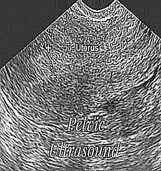
It is carried out by inserting a sensor into the vagina up to 12 centimeters long, the diameter of the device is 3 centimeters. Such a diagnosis is carried out to detect early pregnancy, uterine diseases and other gynecological problems. Conducting a transvaginal examination involves the following steps: a woman undresses to the waist, removing the lower part of her clothing. The patient is laid down on a couch with legs bent at the knees and spread apart on the sides.
The specialist puts a condom on the sensor of the device, lubricates it with a special gel and gently inserts it into the vagina. The helium agent is a conductor between the integument of the body and the sensor. Thanks to the sensor, the visibility of the internal organs is displayed on the monitor of the device.
If the transducer is inserted correctly and slowly, the woman does not feel any pain. The procedure lasts for five minutes.
Transabdominal
The study is carried out by the method of a directed ultrasonic wave through the wall of the peritoneum. Thanks to this diagnosis, it is possible to consider not only a certain organ, but also those adjacent to it. As a result, the specialist gets a clear picture of the organs in the small pelvis.Based on the results of the diagnosis, the specialist makes a diagnosis and prescribes therapeutic therapy.
The transabdomenal method is performed in a supine position (on the back). During the diagnosis, the specialist runs the sensor along the patient's abdominal cavity. In this case, a specific organ is examined. Before diagnostics with a sensor, a helium mass is applied to the patient's abdomen.
Transrectal
This technique is intended for the study of male reproductive, urinary and genital organs. With the help of ultrasound, you can examine the ureter, the prostate gland with seminal vesicles. This study can be performed by both men and women.
The patient should not wear underwear. The patient is examined in a supine position on the left side, with knees bent and pressed more firmly to the chest. The sensor is lubricated with a special agent and inserted into the rectum. The sessions do not cause discomfort or pain. Sometimes, during a session, a specialized specialist can take tissue material for a biopsy, if, of course, such appointments take place.
According to the results of any method of ultrasound, it is possible to study a number of indicators: shape, size, structure, location of the urinary and genital organs.


