Home >
Erectile Dysfunction >
MRI examination of the prostate
MRI examination of the prostate

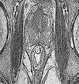
MRI of the prostate gland allows you to detect tumor formation at an early stage, specifying the type and specificity of the pathological process.
Urologists often prescribe a non-invasive technique to study pathological changes in the prostate.
When is an MRI scan of the male gland indicated
Tomography is performed to detect various diseases of the prostate:
- anomalous structure;
- gland growth;
- a type of prostatitis;
- abscess;
- adenoma;
- cancer;
- hyperplasia.
Magnetic resonance imaging is mainly prescribed to identify the oncological process in the male gland, manifestations of metastasis, its location, when choosing an effective therapy for the oncological process.
What types of MRI are there
The male organ is small in size and is located at a considerable depth, therefore, it is not always possible to obtain information by only one effect of a magnetic field. Additional manipulations may be required to increase the information content. MRI examinations are classified according to classifications.
Classic exploration
No additional manipulation. The patient lies down on a special movable surface that slides into a cylindrical structure surrounded by a magnetic field. If it is necessary to fix the patient in a statistical position with a pillow or straps (for immobility), in another version, distorted results may be obtained. The cost of such an examination is expensive.
Contrast MRI
This MRI of the prostate is performed by injecting contrast medium through an intravenous injection. This gives good visibility on the monitor of the device. This examination allows you to see healthy and diseased tissue. An appointment to conduct a contrast study is accepted by a specialized specialist if men have suspicions of an oncological process of the prostate gland.
Preparing for this kind of MRI is a little different than usual. The man undergoes tests to determine the sensitivity to the staining injection. When no problems are found, a cleansing enema is done the day before the examination before going to bed. Food should not be consumed until the research session. MRI of the prostate with contrast is not performed, if the patient has an intolerance of the body to the staining substance, then another type of examination of the state of the organ is chosen.
Before performing the resonant procedure, the doctor injects a contrast fluid through the vein. The further technique of the procedure is no different from the traditional MRI examination.
MRI study using an endorectal coil
This computer procedure is carried out as follows: a thin, flexible hose, covered with a latex sheath, is introduced to a man. Since this wire is close to the organ, an additional magnetic field is created. This method is considered more accurate for assessing the state of the organ. Before examining the state of the organ, an enema is done per day, if the patient suffers from constipation, then it is recommended to take a laxative medication.
Multiparamatic MRI
This type of examination for prostate cancer allows you to combine all types of tomography. Thanks to this study, it is possible to recognize the oncological process even at the earliest stage. This method eliminates any medical error in the diagnosis. With this method of research, the exact localization is determined with the appearance of growths of neoplasms.
After such a diagnosis, the patient does not need a biopsy, which allows the patient to avoid unnecessary punctures.
How patients should prepare for an MRI scan
MRI examination of the prostate, which is prescribed by the attending specialist. He gives the man recommendations for preparatory manipulations, taking into account the clinical picture and physical characteristics.
However, there are a number of mandatory rules:
- for two or even three days, the patient should not consume gas-forming products (cabbage, beans, mushrooms, celery, raw onions, carrots, apples, raisins, pears, kvass, carbonated water, bananas, watermelon, dairy products, flour) ...
- the day before the study, the patient must take charcoal (activated), at the rate of 2 tablets for each kilogram of weight;
- food should not be eaten 6 hours before the session;
- No-shpa is taken an hour before the session;
For whom is MRI of the male gland contraindicated
Like any procedure, MRI has its own contraindications, the presence of which is prohibited to conduct such a study:
- implantation in the heart muscle;
- large metalized prostheses;
- electronic or metal hearing aid implants;
- the presence of clips (for cerebral aneurysm) made of metal material;
Indirect contraindications include:
- severe pathological processes of the heart (acute heart failure);
- psycho-emotional disorders;
- tattoos containing metal paint;
- overweight, obesity;
- heart valve prostheses;
- pacemakers;
- insulin pumps;
- claustrophobic.
Pros and cons of MRI research
There are positive and negative aspects of magnetic resonance imaging of the prostate. The advantages include the safe use of modern, high-quality tomographs, which makes it possible to preserve the integrity of the skin. This method of studying the state of the organ guarantees the fact of protection from ionizing radiation. With the help of this type of research, specialists can obtain accurate detailed information about the image of an organ with all its flaws.
MRI allows you to examine the chemical structure of the male gland. The research is carried out for half an hour. Magnetic resonance imaging, practically does not manifest an allergic reaction. For example, on contrast liquid, if such a phenomenon still occurs, then this is noted in extremely rare cases.
Contraindications for this type of study of the prostate gland in men include the cost of the procedure. Not a cheap pleasure.
Alternative Prostate Diagnosis
When patients do not have the opportunity to conduct a tomographic study of the prostate, there are alternative research methods:
For each patient, the method of research is selected by the doctor based on the general state of health, clinical picture of pathology, age, financial capabilities of the patient. If a specialist comes to the conclusion of computed tomography, then this means that he has a lot of doubts about the correct diagnosis.


