Home >
Erectile Dysfunction >
Ultrasound of the prostate
Ultrasound of the prostate

Ultrasound of the prostate gland in men, allows you to determine the state of the glandular organ, on which the health of a man depends in full. The prostate gland should be examined annually for prophylaxis, especially for those of the stronger sex who are over 45 years old. Doctors recommend an annual Ultrasound examination of the glandular organ of men.
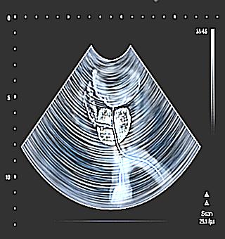
Ultrasound examination of the prostate is a safe, high-frequency diagnosis using sound waves. The picture of the organ is displayed on the monitor screen, where the state of the organ is seen in real time. Using the image, a specialist can examine an organ, determine its structure, structure, size, presence of tumors, and more. Sound examinations are completely safe for human health. Therefore, this diagnosis can be performed as many times as necessary.
When ultrasound diagnostics are scheduled
Sound Research can be used without waiting for health problems or disturbing symptoms. The prostate gland is susceptible to various negative influences from the environment and a man's lifestyle.
The procedure is shown to be carried out for a number of indications, among which there are problems:
- Difficulty emptying the bladder, with a residual feeling of incomplete emptying;
- problem conception, probable infertility, impaired potency;
- induration in the area of the prostate gland;
- pain syndrome in the pubis, prostate, perineum;
- negative results of plasma, semen, urine tests;
- acute kidney or liver disease (failure);
- ultrasound is performed to prevent the condition of the pelvic organs;
- in the complex diagnosis of urinary and genital organs.
How ultrasound diagnostics works
How is prostate ultrasound performed? There are two methods of ultrasound diagnostics. One diagnosis can be made through the peritoneal wall (abdominal), the other - transurectally (through the rectum). Preparation is required for both examinations. Transrectal diagnosis is also called TRUS. Both sessions are performed on an empty stomach, and transrectal examination requires dietary restrictions.
Preparing for Surface Diagnostics
Preparing for a transabdominal examination is easy. Before carrying out the inspection, you must take about a liter of water. The liquid is used one hour before the examination. With a full urinary tract, the specialist has more opportunities to examine the organ. Preparation for the diagnosis of ultrasound begins two or three days before the session. To do this, the doctor recommends trying to remove gases from the body.
Before conducting an ultrasound scan, the patient should drink activated charcoal for three days, which will help remove gases. The transducer of the ultrasound machine clearly sees the changes in the organ through the liquid, but the gases prevent the apparatus from diagnosing the organ.
For the decoding to be accurate, the bladder must be filled to the point of a desire to urinate, but not critically. Thanks to such a diagnosis, the man cannot tolerate psychological experiences, which cannot be said about the study by the transurethral method. However, an abdominal examination will not give the desired results if the man has excess body weight, since there is a large fat layer between the sensor and the organ. For example, it may be possible to study the size of the prostate gland, but it will not be possible to detect small structural changes.
Making an ultrasound is quite simple. The patient lies down on a couch (supine position), undressed to the waist. The doctor applies a special gel to the peritoneum and diagnoses the organs with the help of a transducer. This method allows you to diagnose the urinary tract, liver, digestive organs, kidneys (lying on its side), prostate, detect stones and more.
Rectal prostate diagnostics
How is ultrasound done? Transrectal examination of the glandular organ is carried out by the method of penetration of the sensor (electronic) into the rectum. This method eliminates the obstruction of the body fat. The screen picture is clearer. Urologists most often prescribe a transrectal examination of the prostate gland.
The positive qualities of this method include:
- non-invasiveness;
- short session time;
- painless;
- no discomfort;
- even the vessels of the prostate can be seen;
- study of all lobes of the organ;
- inspect the structure and more.
When a patient has problems with the anal canal (cracks, inflammation, hemorrhoids), the doctor may postpone the examination for a while to exclude an acute condition.
How is rectal ultrasound done? The man lies down in a position on his side, with the knees bent and tightly pressed against the abdominal cavity.The probe is inserted into the rectum. The small size of the sensor allows the patient not to feel any special discomfort. The procedure is carried out within a quarter of an hour. To improve glide, the sensor is protected by a special condom and lubricant, which allows the device to glide faster.
Preparatory manipulations for the transrectal examination
For transrectal ultrasound of the prostate, the patient needs special training. Before diagnosing with this method, the patient maintains a special diet. For this, in three days, all products that have a defiant effect on flatulence or tight stools are excluded. The patient is not recommended to consume dairy products, rye and black bread, fruit (sour), grapes, legumes. Soups, water-based cereals should be present in the diet.
In the evening, before the morning examination, a cleansing enema is performed or using suppositories.
What pathological processes of the urinary and genital area are determined by ultrasound
What does an ultrasound of the prostate show?
- Development of adenoma (benign neoplasm).
- Oncological processes.
- Erectile dysfunction.
- Cystosis and polyps.
- Examination of urea for urolithiasis.
In a healthy man, the organ should have smooth organ contours, with symmetrical and clear edges. On examination by a doctor, the shape, structure, size, uniformity are determined, and calculations are made. Based on the test results, the mass and volume of the prostate gland are calculated using a special formula. A normal prostate should not be more than 27 cc.
When the development of an oncological process is suspected, the doctor prescribes a tissue examination for a biopsy. This manipulation is performed with a special needle, while the patient does not have an unpleasant sensation, since the rectum has a low sensitivity to pain.
Conducting an ultrasound scan cannot be ignored, especially for older and middle-aged men. With the help of such an examination, pathological processes are detected at the initial level of the disease, which makes it possible to start effective therapy in a timely manner, avoiding serious consequences.


